Understanding the Mexican Red Knee Tarantula Anatomy
The Mexican Red Knee Tarantula (Brachypelma hamorii), a captivating creature, is a popular choice for pet owners and a fascinating subject for arachnologists. Understanding the intricate anatomy of these tarantulas is key to providing proper care and appreciating their unique adaptations. This guide delves into the various aspects of their anatomy, from the protective exoskeleton to the internal organs that keep them functioning. By exploring the structure and function of each part, you’ll gain a deeper understanding of what makes these spiders so special. This knowledge is not only helpful for enthusiasts but also crucial for anyone considering keeping a Mexican Red Knee Tarantula as a pet, ensuring their health and well-being. The more you know about these amazing creatures, the more you will appreciate their beauty and complexity. This comprehensive guide will walk you through each component, helping you understand the intricacies of the Mexican Red Knee Tarantula’s anatomy.
Exoskeleton The Protective Shell
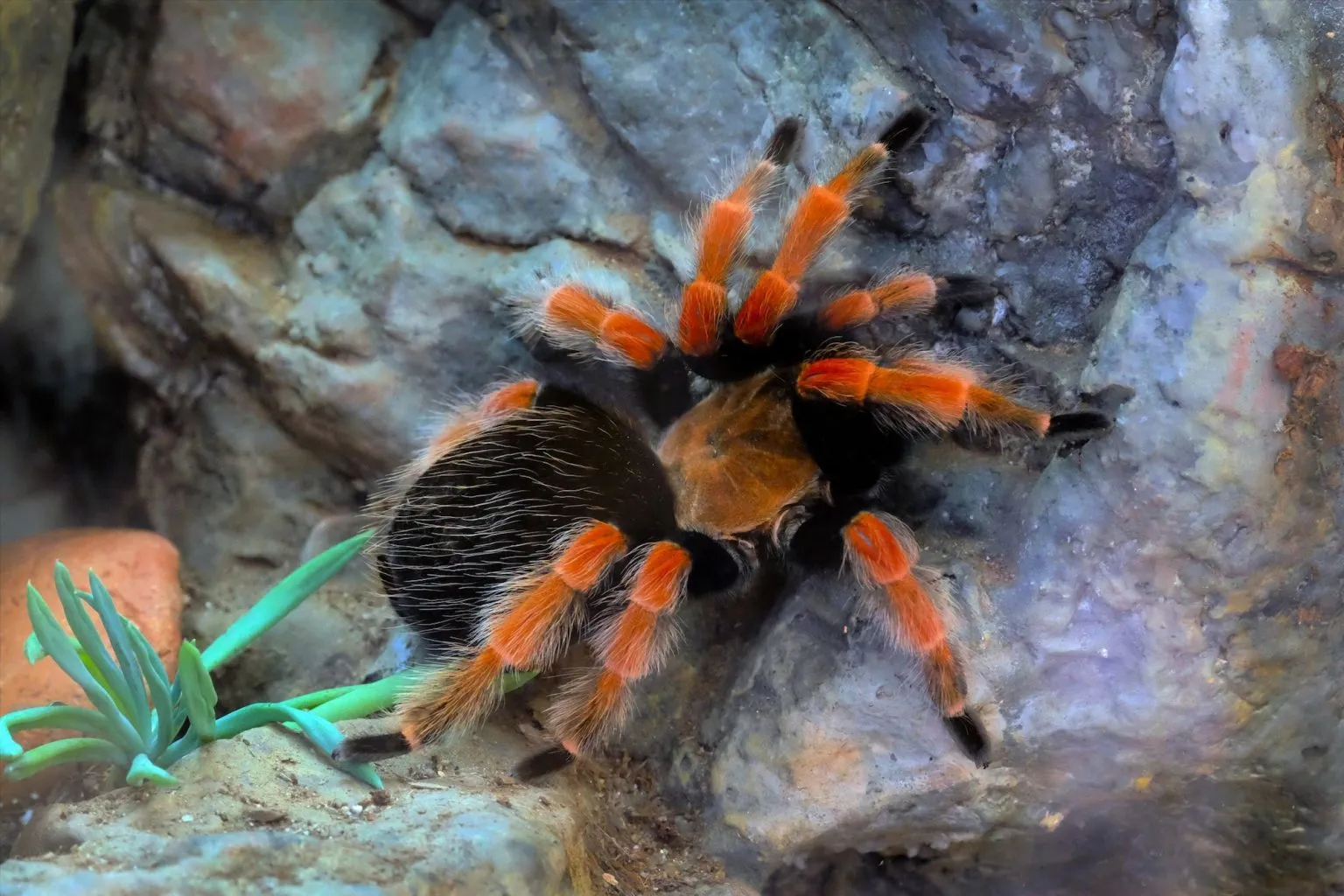
The exoskeleton is the defining feature of all arthropods, and in the Mexican Red Knee Tarantula, it serves as a robust protective armor. Unlike vertebrates with internal skeletons, tarantulas have an external skeleton made primarily of chitin, a tough, flexible polysaccharide. This exoskeleton protects the tarantula from predators, physical damage, and water loss, acting as a first line of defense against the outside world. The exoskeleton’s rigidity also provides structural support for the tarantula’s muscles, enabling movement. The color and patterns on the exoskeleton, particularly the vibrant red-orange bands on the legs, serve both for camouflage and mate recognition. Understanding the exoskeleton’s role is crucial because it influences how the tarantula interacts with its environment and its vulnerabilities during molting.
Structure of the Exoskeleton

The exoskeleton is not a single piece of armor but rather a complex structure composed of several layers. The outermost layer, the epicuticle, is waxy and waterproof, preventing the tarantula from drying out. Beneath this is the procuticle, which is the thickest layer and provides the main structural support. The procuticle itself is made up of two layers the outer exocuticle, which is hardened and the inner endocuticle, which is more flexible. These layers work together to give the exoskeleton both strength and flexibility. The exoskeleton also has different thicknesses in different parts of the body the abdomen is more flexible to allow for expansion after a meal, while the cephalothorax is more rigid to protect vital organs. The structure of the exoskeleton is critical for the tarantula’s survival, providing protection, support, and a barrier against the external environment. This complex architecture enables the tarantula to thrive in its habitat.
Shedding the Exoskeleton Ecdysis
Because the exoskeleton is rigid, the tarantula must shed it to grow. This process, called ecdysis or molting, is a crucial part of the tarantula’s life cycle. Before molting, the tarantula creates a new, soft exoskeleton beneath the old one. Enzymes are then secreted to detach the old exoskeleton. The tarantula absorbs water and swells, causing the old exoskeleton to split open, usually along the cephalothorax. The tarantula then wriggles out of the old exoskeleton, leaving behind a perfect replica of itself, including the lining of the gut and the book lungs. After molting, the tarantula is vulnerable as its new exoskeleton is soft and takes time to harden. During this period, the tarantula is more susceptible to injury. Molting allows the tarantula to grow larger, regenerate lost limbs, and remove parasites. The frequency of molting decreases as the tarantula matures, but it remains a vital process throughout its life.
Body Segmentation
The body of the Mexican Red Knee Tarantula, like all spiders, is divided into two main sections cephalothorax (prosoma) and abdomen (opisthosoma). This segmentation is a fundamental characteristic of arachnids and influences the spider’s functionality. The cephalothorax is the fused head and chest region, where the eyes, mouthparts, legs, and other sensory organs are located. The abdomen contains the digestive system, reproductive organs, and book lungs. The connection between the cephalothorax and abdomen is a narrow pedicel, which allows for flexibility and movement. Each section of the body serves a specific purpose, contributing to the spider’s survival and ability to thrive in its environment. Understanding body segmentation is critical for appreciating the spider’s body plan and how it functions. The arrangement of these two sections makes the tarantula a highly specialized predator.
The Cephalothorax Head and Thorax Combined

The cephalothorax is the control center of the tarantula. It’s where the tarantula’s head and thorax are merged. This fused region houses the eyes, mouthparts (chelicerae and pedipalps), and all eight legs. The cephalothorax is protected by a hardened carapace, which is a part of the exoskeleton. The arrangement and structure of the cephalothorax are designed to support the spider’s feeding, movement, and sensory functions. The eyes, usually eight in number, provide a limited view of the surroundings, detecting light and movement. The chelicerae, equipped with fangs, are used for injecting venom and manipulating prey. The pedipalps are used for sensing, feeding, and in males, for reproduction. The cephalothorax is a critical region for the tarantula’s survival, containing all the essential structures needed for feeding, movement, and sensory perception.
Eyes and Mouthparts
Mexican Red Knee Tarantulas have eight eyes, arranged in two rows. These eyes are not as advanced as those of some other animals, but they allow the tarantula to detect light, shadow, and movement. This is essential for hunting and avoiding predators. The mouthparts consist of chelicerae, which contain fangs, and pedipalps. The chelicerae are used to inject venom to subdue prey and manipulate food. The pedipalps, located near the mouth, are used for sensing the environment, manipulating food, and in the case of mature males, for transferring sperm. The combination of eyes and mouthparts are a vital part of their survival. The fangs are designed to pierce the exoskeleton of prey, while the pedipalps assist in the feeding process and provide additional sensory information.
The Abdomen
The abdomen is the softer, more flexible posterior section of the tarantula’s body, connected to the cephalothorax by a narrow pedicel. This section houses vital internal organs, including the digestive system, reproductive organs, book lungs, and the heart. The abdomen’s flexibility is important, particularly after feeding, as it allows the tarantula to expand to accommodate a large meal. The abdomen is also where the spinnerets are located, which are used to produce silk. The abdomen is covered in sensory hairs that aid in detecting vibrations and movements. The abdominal structure is critical to the tarantula’s overall health, housing the systems that help it to digest food, breathe, reproduce, and maintain internal balance. This section is also where they store reserve food, making it essential for their survival.
Book Lungs

Book lungs are the primary respiratory organs of the Mexican Red Knee Tarantula, located in the abdomen. These structures get their name from their resemblance to the pages of a book. Each book lung consists of numerous thin, leaf-like lamellae filled with hemolymph (the spider’s version of blood), separated by air spaces. Oxygen diffuses into the hemolymph across the thin lamellae, and carbon dioxide diffuses out. Book lungs provide a large surface area for gas exchange, enabling the tarantula to breathe effectively. The number and size of book lungs can vary, affecting the tarantula’s activity level. The efficiency of these book lungs is crucial for the tarantula’s energy production and overall health. Book lungs are vital in providing oxygen to the spider, essential for all its bodily functions.
Spinnerets
Spinnerets are silk-producing organs located at the posterior end of the tarantula’s abdomen. These structures are responsible for producing silk, which the tarantula uses for various purposes, including creating webs, lining burrows, and wrapping prey. Each spinneret is composed of numerous spigots, which secrete silk fibers. The tarantula can control the flow and texture of the silk, using it to construct different types of structures. The silk is incredibly strong and flexible, making it perfect for trapping prey. The ability to produce silk is critical for the tarantula’s hunting success and shelter construction. Spinnerets are one of the most important features of a tarantula, enabling it to build its home, catch its food, and provide a safe environment.
Legs and Movement
The Mexican Red Knee Tarantula has eight legs, each consisting of seven segments coxa, trochanter, femur, patella, tibia, metatarsus, and tarsus. These segments are connected by flexible joints, allowing the tarantula to move with agility. The legs are covered in sensory hairs (setae) that help the tarantula detect vibrations, and also have claws on the tarsi to grip surfaces. The legs are also covered with hairs that serve as a means to climb and also sense the environment. The legs of the Mexican Red Knee Tarantula have distinct characteristics. The vibrant red-orange bands on the legs are a key identifying feature. The legs play a vital role in locomotion, hunting, and sensing the environment. The specialized structure of the legs, combined with the sensory hairs and claws, enables the tarantula to move across various terrains with ease, hunt efficiently, and sense the world around it.
Sensory Hairs

Sensory hairs, also known as setae, cover the entire body of the Mexican Red Knee Tarantula, including the legs, body, and mouthparts. These hairs play a crucial role in the spider’s sensory perception. There are different types of sensory hairs, each with a specific function. Some hairs detect vibrations, helping the tarantula sense the approach of prey or predators. Other hairs are sensitive to air currents, providing information about the environment. The sensory hairs also help the tarantula to navigate and perceive its surroundings. The sensory hairs enable the tarantula to live a very sophisticated life, offering an enhanced perception of its habitat. They are the key to their survival in their environment, allowing them to detect the smallest changes around them.
Internal Organs
The internal organs of the Mexican Red Knee Tarantula are located primarily within the abdomen, and are essential for the spider’s survival. These organs are critical for maintaining the tarantula’s life functions. The digestive system processes food, the circulatory system transports nutrients and oxygen, and the nervous system coordinates the spider’s activities. The internal organs are often delicate and protected by the tough exoskeleton. Understanding these organs is fundamental for comprehending the overall health of these fascinating creatures. The internal organs of the tarantula function in a coordinated manner, supporting the spider’s daily activities.
Digestive System
The digestive system of the Mexican Red Knee Tarantula is adapted for consuming liquid food. The tarantula injects digestive enzymes into its prey, breaking down the tissues into a liquid form. This pre-digested food is then sucked up through the mouth. The digestive system includes a pharynx, esophagus, stomach, and intestines. The stomach and intestines are responsible for absorbing nutrients. The waste products are eliminated through the anus. The digestive system is one of the most important systems for the tarantula. The process of breaking down prey externally allows the tarantula to consume a wide range of food sources. The digestive system is a highly specialized and efficient system that allows the tarantula to thrive in its environment. Understanding the digestive system is vital for understanding the tarantula’s feeding habits and health.
Circulatory System

The circulatory system of the Mexican Red Knee Tarantula is an open system, meaning that the blood (hemolymph) does not circulate entirely within vessels. The heart, located in the abdomen, pumps hemolymph through arteries, which then empty into body cavities, bathing the organs directly. The hemolymph transports nutrients, oxygen, and waste products. The hemolymph flows back into the heart through openings called ostia. The circulatory system is responsible for the transport of vital substances throughout the tarantula’s body. Oxygen is delivered to the tissues by the hemolymph, and carbon dioxide is carried away. The open circulatory system works efficiently for the spider, allowing it to survive in its habitat. The heart’s pumping action and the hemolymph’s function are crucial for maintaining the spider’s health and functionality.
Nervous System
The nervous system of the Mexican Red Knee Tarantula controls its movements, senses, and behaviors. The main part of the nervous system is the central nervous system, which consists of a brain (a cluster of ganglia in the cephalothorax) and a ventral nerve cord running along the underside of the body. The brain receives and processes sensory information and controls the tarantula’s responses. Nerves branch out from the brain and ventral nerve cord to all parts of the body, allowing the tarantula to sense its environment and coordinate its movements. The nervous system enables the tarantula to react to its surroundings. Sensory information is processed, and appropriate responses are initiated. The tarantula’s nervous system allows it to hunt, avoid predators, and perform other vital functions.
Reproductive Anatomy
The reproductive anatomy of the Mexican Red Knee Tarantula is different in males and females, playing a crucial role in the species’ reproduction. Understanding the reproductive structures is key to understanding their mating behavior. During reproduction, male tarantulas transfer sperm to the female, who then lays eggs. The process is delicate and intricate, and the reproductive anatomy reflects this complexity. The internal and external reproductive parts are specialized for this process. The female’s anatomy allows her to receive, store, and fertilize the eggs, while the male’s anatomy is adapted for delivering sperm. These structures are vital to ensure the survival of the species and make the reproductive phase one of the most fascinating aspects of tarantula biology.
Male Reproductive System

The male Mexican Red Knee Tarantula has a relatively simple reproductive system. Males have two pedipalps, which they use for sperm transfer. Before mating, the male creates a sperm web on which he deposits his sperm. He then loads his pedipalps with the sperm. During mating, he inserts his pedipalps into the female’s epigastric furrow to transfer the sperm. The pedipalps have a bulb-like structure containing the sperm. The male’s mating behavior, including the use of his pedipalps, is critical to the reproduction of the species. The pedipalps are essential for sperm transfer. The male’s reproductive anatomy is specifically adapted to this purpose. The efficiency of this system is crucial to the survival of the species. The sperm transfer process is a fascinating aspect of the male tarantula’s life cycle.
Female Reproductive System
The female Mexican Red Knee Tarantula has an internal reproductive system. She has an epigastric furrow on her abdomen, where she receives sperm during mating. The female stores the sperm within her spermathecae, internal storage sacs. The eggs are fertilized internally. After fertilization, the female produces an egg sac made of silk, in which she deposits her eggs. She guards the egg sac until the spiderlings hatch. The female’s reproductive anatomy is designed to receive, store, and fertilize eggs. The internal system is highly specialized to support the development of the offspring. Her care of the egg sac is essential for the young spiders’ survival. The female’s reproductive process is a complex and fascinating process, critical to the continuation of the species.